Escape Strategies When Using Simple External Skeletal Fixators
by IMEX Veterinary
The previous three issues of the IMEX Update introduced the SK ESF System, encouraged users to construct simpler frame geometries using the proper sized SK fixator, and provided case examples of successful fracture management utilizing simple ESF frames. This issue will review the design philosophy of the SK ESF System and the economic and biologic advantages of simple, half-pin based frames. More importantly, this issue will introduce the concept of taking advantage of the fact that certain fixation pin sizes can be effectively gripped by two different SK clamp sizes as a handy method of staged disassembly when one begins with a simple ESF frame.
A historical look at veterinary ESF reminds us that the original Kirschner-Ehmer (K-E) external fixator dominated the veterinary market for decades and was originally marketed as a half-pin splint or a Type I-a construct utilizing smooth fixation pins. The KE device used in such a fashion did not perform well and as a result, external skeletal fixation generally fell out of favor in veterinary practice. Fortunately, veterinary surgeons and researchers were able to produce improved clinical results by incorporating threaded pins, constructing more complex ESF frames based on full-pins, and stacking (augmenting) external bars. These mechanically improved frames greatly reduced patient morbidity as compared to simple half-pin frames as originally attempted, but resulted in expensive frames that were time consuming and difficult to apply. Veterinarians then began looking for ESF devices and instruments that would simplify application of complex (multilateral, multiplanar) ESF constructs and simplify use of positive threaded ESF pins. Acrylic frames, expensive aiming devices, and bar stacking devices were developed to facilitate the construction of complex frames.
The SK ESF clamp was designed to:
These design features of the SK ESF clamp proved very popular with surgeons beta testing the SK clamp prototypes and truly simplified construction of complex frames and use of modern ESF pins. However, engineers were not happy with merely simplifying the construction of complex frames, but wondered why complex frames were necessary and if it would be possible to design an external skeletal fixator that would perform well using simple, half-pin based frame geometries. Mechanical testing of Type I-a frames demonstrated that the traditional KE external bar is the weak link of the frame. Bar deformation is usually not permanent, but results in premature pin loosening and poor limb function (morbidity). Complex frames, full-pins, and stacked external bars are utilized to overcome the inadequate bar strength of KE and other weak bar devices.
FIGURES 1 and 2 | Mechanical testing of Type I-a frames demonstrates that the traditional external rod is the weak link. Bending of the rod is not usually permanent, but results in premature pin loosening and poor limb function. Complex frames can hide weak rods. Strong rods are mandatory to achieve simple but effective frames. (Bronson, D.G., Ross, J.D., Welch, R.D., Proceedings of Veterinary Orthopedic Society Annual Meeting, 1999)
Comminuted, Midshaft Fracture of the Radius/Ulna
4-year-old, male Labrador, 36 kg

FIGURE 4 | Demonstrating technique - destabilization was made easier by removing half the frame and adding small SK components before removing the other half of the large SK, Type I-b frame.

FIGURE 5 | Resulting 4-pin, small SK, Type I-a frame. Early healing supports this aggressive destabilization strategy.
Tibial Fracture
1-year-old, intact, male German Shepherd, 40 kg

FIGURE 7 | 6-week radiograph demonstrating excellent fracture healing. The fixator was removed at this time.

FIGURE 8 | Gross photo of patient 18 hours postoperatively.
Grade I, Open Tibial Fracture
18-month-old, intact, male Airedale
Fortunately, engineers were able to convince veterinary surgeons that the most powerful way to simplify the ESF method lies not with instrumentation and clamps that simplify construction of complex frames, but in elimination of the weak link of simple frame constructs. Specifically, the SK ESF System replaced the historically utilized 3.2 mm and 4.8 mm KE external rods with larger diameter (stronger) external rods while retaining all of the user-friendly features of the prototype SK ESF clamp previously outlined. For 6 years IMEX customers have enjoyed the user-friendly clamp design of the SK ESF System. Many, but not all, have taken advantage of rigid SK external bars to simplify frame geometries and several cases are shown again to demonstrate the capability of SK ESF frames.
Historically, as veterinary surgeons evolved from simple KE frames to frames utilizing multiple full-pins in each major bone fragment, the axial stiffness of the frames increased dramatically. In fact, regardless of device or external bar, frames utilizing multiple full-pins are all very stiff under axial loading. Since this high level of axial stiffness will sometimes slow bone healing, it became popular to convert complex frames to less stiff frames as early stages of healing occurred. This sequential frame disassembly may be done in one step or in several and is termed staged disassembly. The planned reduction of fixator rigidity transfers more of the load bearing forces across the bone, stimulating callus maturation and the later stages of bone healing. Common examples of converting a complex frame to a simpler, less rigid frame include: conversion of a Type III frame to a Type II frame, conversion of a Type II frame to a less complex Type II frame, or conversion of a Type II frame to a Type I frame. Other examples include conversion of a Type I-b frame to a Type I-a frame or removal of articulations between different components of a bi-planar frame.
If one begins with a simple Type I-a frame the previously listed options for staged disassembly are not applicable, however, two alternate strategies can be utilized. First, reduction of pin number is an option with the SK ESF System that is often considered risky with KE and KE-like devices. If early callus formation is occurring, reduction in pin number - especially removal of the pins on each side of the fracture site resulting in increased external bar working length - is potentially a safe method of staged disassembly. This assumes adequate initial pin number and the presence of soft callus that creates some stability to the fracture zone. Alternatively, if reduction in pin number might jeopardize adequate pin bone interfaces, substituting a smaller, more flexible external rod for a larger, stronger one becomes a very attractive option to decrease the stiffness of a Type I frame (e.g. removal of large SK clamps and a 9.5 mm carbon fiber composite rod and replacing them with small SK clamps and a 6.3 mm titanium rod). While not truly a disassembly, rod downsizing does achieve the purpose of transferring a greater percentage of the load bearing forces back to the bone and across the healing callus. A variation of this concept when utilizing the small SK device with 6.3mm titanium rods is to replace the titanium rod with the less rigid carbon fiber composite rod of the same diameter. While not “downsizing” the connecting rod, this method does achieve a similar planned decrease in rigidity and might be useful in dogs where initial construction utilized size medium fixation pins (see Bending Stiffness Comparison of External Rods).

FIGURE 11 | Bending Stiffness Comparison of External Rods
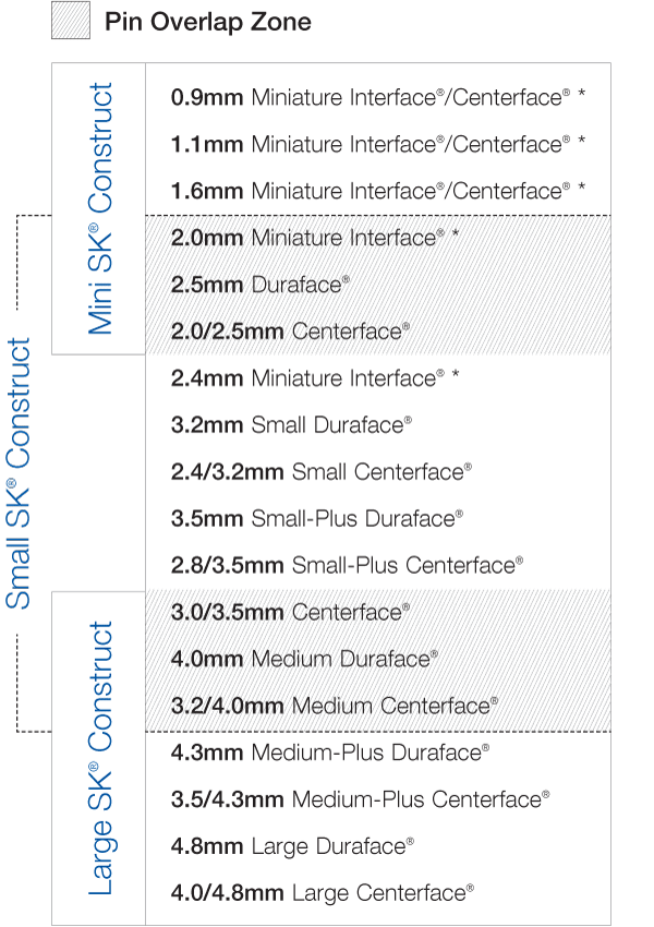
Since each SK clamp is designed to grip a wide range of pin diameters, and there is an overlap zone between the different sizes of fixation pins gripped by the different SK clamp sizes, it is frequently possible to construct the initial fixator with the larger clamps and rods and at about six weeks replace these components with those one size smaller. This wide range of pin shank diameters that can effectively be gripped with the SK clamp makes utilization of “overlapping pin zones” particularly useful with the SK device. The downstaging disassembly chart summarizes the pin sizes that each of the three SK ESF devices can utilize in a through-the-clamp fashion, as well as the bar/clamp downstaging potential of various pins used for initial frame construction using the large or small SK fixator.

* Miniature Interface and miniature Centerface pins continue to be used according to their smooth shaft dimensions. Standard size Interface and Centerface naming convention now includes both shaft and thread diameter.
FIGURE 12 | Pin Overlap Zone
Not all fixator frames will require staged disassembly. In particular, young patients tend to produce bony callus rapidly and much less often need or benefit from staged disassembly (see case 2). All patients will benefit from early fracture stability which promotes fracture zone debridement, revascularization, and early callus formation. Only after these stages occur will the potential benefits of decreased rigidity become pertinent. With several options for converting more complex frames to less complex frames, or downstaging larger, stronger bars to smaller, less rigid ones; it is prudent to consider use of the stronger choice initially with a staged exit strategy available later if so needed. In skeletally mature canine patients, the optimal time period for initiating staged disassembly appears to be at about 6 weeks after surgery.


