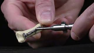
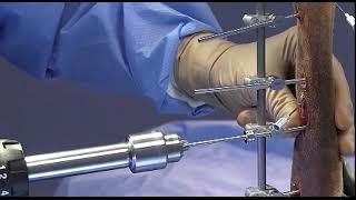
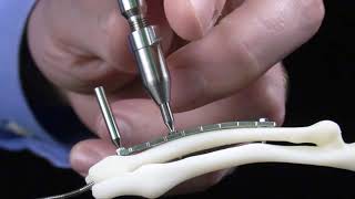
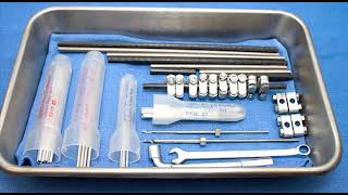
Using and Storing CrossCut Wedge Osteotomy Guides
Correction of angular limb deformities in small animal patients can be challenging and begins with detailed planning of the correction which often involves a closing wedge ostectomy.
Read more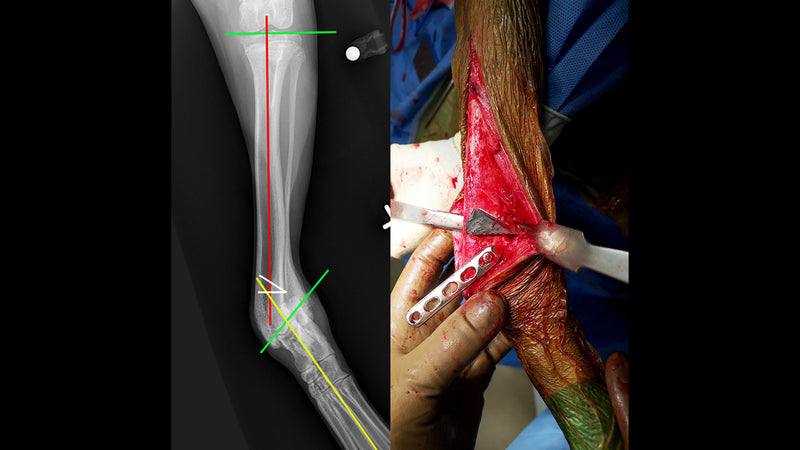
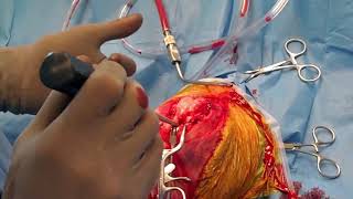
Stabilization of Coxofemoral Luxation Using the Toggle Pin Method
There are a number of popular surgical methods for maintaining reduction of coxofemoral luxations - use of the toggle pin method, ilio-femoral sutures to limit external rotation of the hip, and caudo-distal transposition of the greater trochanter.
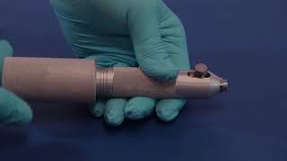
Read moreModern ESF Pin Terminology
For many years, external fixation pin use centered around the Kirschner-Ehmer (KE) external fixation device and newer modifications of it. This resulted in potentially confusing pin terminology...
Read moreFirst Major Improvement in ESF Pin Technology
IMEX Duraface half-pins demonstrate a 55% average increase in stiffness and a 3.7 fold increase in cyclic fatigue life compared to current positive-profile pins without an increase in cost. These mechanical improvements can...
Read more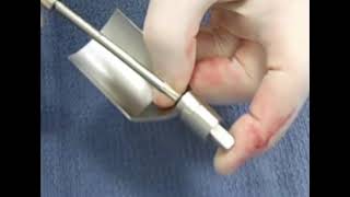
Hybrid ESF Fracture Repair of Short Distal or Proximal Segments
Hybrid external skeletal fixation (HESF) is a valuable tool for repair of fractures with a short distal or proximal segment. Short, juxta-articular fracture segments are often difficult...
Read more